
Individual neurons in both hemispheres of the murine model have been found to exhibit elaborate branching patterns similar to embroidery, produce an expansive brain atlas, and often release spindle fibres that are around half a metre in length.
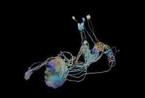
This 3-D map of the mice psychological makeup is the most complete effort to date at tracing the connections between the brain’s various regions. The map, which is the product of a dedicated group of scientists somewhere at Genelia Research Campus, corrects the orientation of over 300 of the over 70 million cells in a murine model. There have been hundreds of prior efforts to trace the journey of a single neuron.
Mind Mapping’s Functional Potentials
“Three hundred synapses is only the commencement,” says biologist Jairam Chandrasekhar, whom heads the Genelia team members, also known as the MouseLight for their attempts to shed light on the connectivity of the rat brain.
There in following months, he and his team aim to discover hundreds more neurons, and they will be making all of their findings public for the benefit of the neuroscience field.
Read Also: Brain Computer Interface: How Close Are We
In October 27, 2017, the team published its most recent dataset along with an analytic device called MouseLight NeuronBrozer. They will present their findings at the next annual meeting of the Society for Neuroscience in Washington, DC.
In-Depth Brain Map Analysis
In search of the research results, Questions will aid in asking and beginning to answer, questions regarding the organisation of neurons and the movement of information throughout the brain. Director of the Genelia project teams and neuroscientist Wyatt Korff deems the new MouseLight dataset to be the biggest of its type. That’s going to alter how neurons are described in books, experts say.
Neurons resemble poached eggs with a tail in most biology textbook illustrations. Speeding signals are sent down the axon of a neuron, and other neurons may take them up at their dendrites. Information zips back and forth between these cells, allowing us to do things like reach for a coffee pot or find our vehicle keys.
According to Chandrasekhar, specialists have known for a while that neurons are overlooked in the textbook approach. However, their findings imply that connections between neurons are typically far more extensive and robust than scientists had previously thought.
Tools for Making Comprehensive Maps of the Brain
Brain connectivity has been extensively mapped by researchers, but obtaining a zoom-in with human limitations in mind is technically challenging. Extending single neurons from one of the millions of photos obtained requires scientists to first expose them within a mouse brain, then turn the brain into thin sheets, and last photograph the sheets using a microscope.
Genelia’s staff attacked the issue by honing their procedures. According to Korff, “from a team scientific point of view, this effort was only doable.” Neuroscientists, anatomists, computer programmers, and software developers were all represented among the project’s researchers.
Before anything else, the researchers introduced a virus into the mouse brain, which can only infect a small number of neurons. The brain is then “cleaned” so that light can better reach the tissue. Finally, 200-micrometer brain slices are closed with a buzzing knife after being subjected to light pulses from a state-of-the-art light microscope. This procedure is continued until a complete copy of the brain has been made.
Neuronbrozer
Using a speedy version of a technique called “two-photon microscopy,” we can spotlight neurons and prevent other focus planes from being illuminated.
This provides the high-quality pictures required for brain mapping studies. Each mind generates over 20 terabytes of information, which is equivalent to roughly 4,000 DVDs. Images are stitched together using advanced algorithms, and then seven trained humans perform the resultant dataset down to the neuron level.
Neuroscientist and regional director of scientific programmes at Genelia Nelson Spryston emphasises the importance of these individuals and the tools they use as essential components of high-quality mapmaking.
Large-Scale Brain Functions and Mapping
Prior to these last two years, it took an individual at least a week, and sometimes more, to follow a neuron as it travelled throughout the brain. He estimates that now each member of the team can monitor around one neuron per day utilising the nerve cell procedures and technology created by the Numerical Computation Group of MouseLight and Genelia.
Spryston adds, “We surpassed 1,000 overall nerves in roughly a year now that our imaging-and-tracing infrastructure is fully operational.” The team’s goal is to reach this level so that they can better understand the information about how neurons transport data in the brain.